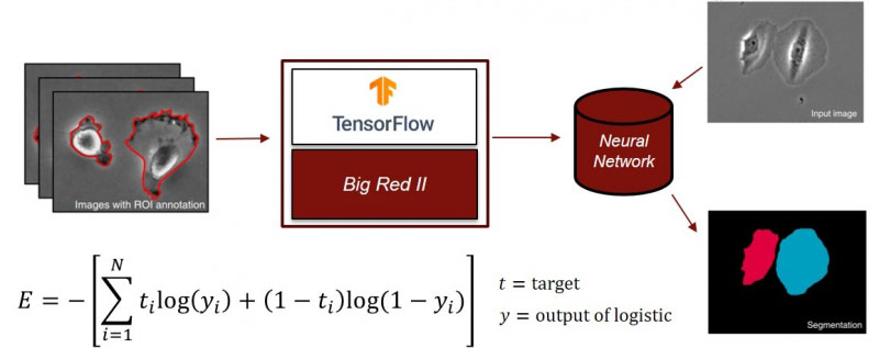
Various computational models have been utilized in our imaging processing/analysis pipeline to automatically recognize and analyze subjects in the image. For example, machine learning and neural networking are used to specifically and correctly segment individual cells in various environments; supervised machine learning is used for single cell phenotyping, and deep learning is used for customized imaging analysis, such as denoise, image registration, and feature extraction.
Computational image analysis: from analytics to informatics

Related publications.
- CSBC/PS-ON Image Analysis Working Group (J. Liu), C. Vizcarra, E. A. Burlingame, C. B. Hug, Y. Goltsev, B. S. White, D. R. Tyson, and A. Sokolov, “A community-based approach to image analysis of cells, tissues and tumors”, Computerized Medical Imaging and Graphics, 95, 102013 (2022).
- Kefer, F. Iqbalb,d, M. Locatelli, J. Lawrimore, M. D. Zhangb, K. Bloom, K. Bonin, P.A. Vidi, and J. Liua, "Performance of deep learning restoration methods for the extraction of particle dynamics in noisy microscopy images" Molecular Biology of the Cell, 32, 903-914 (2021).

